Probably everyone has heard of at least one or the other, maybe even had it done, but doesn’t necessarily know the difference between the two.
Both X-rays and CT scans are often used in healthcare to get a better picture (literally) of a certain area of the body in a non-invasive way.
Below are the specifications of both procedures and the differences between the two. At Evergreen Dental, both dental x-rays and CT scans are available.
Both X-rays and CT scans use low doses of radiation. This may sound scary or even disturbing to most people, but usually the amount of radiation you are exposed to during an X-ray scan is equivalent to between a few days and a few years of exposure to natural radiation from the environment. Although there is no magic number for how many x-rays are safe to take in a year, the American College of Radiology recommends limiting lifetime diagnostic radiation exposure to 100 mSv (a millisievert is a radiation protection unit), which is equivalent to about 10,000 chest x-rays but only 25 chest CT scans.
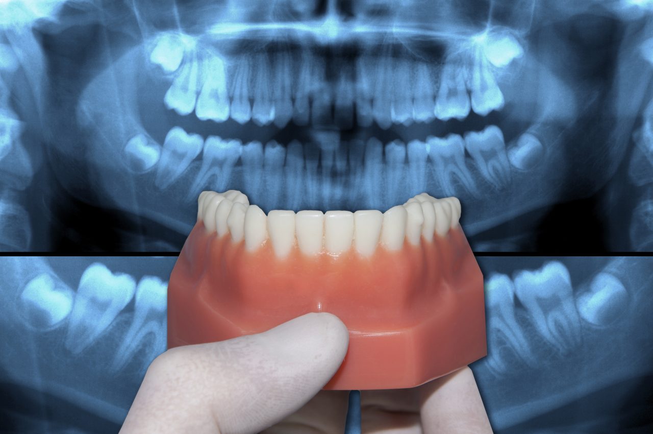
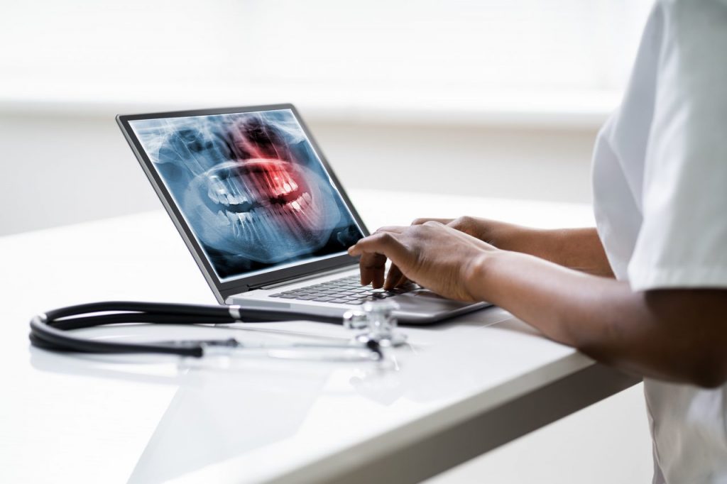
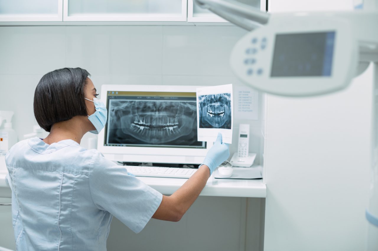
Dental X-rays are used to take two-dimensional images of your teeth. They give your dentist valuable information about your teeth and gums, and help him or her to make the best treatment plan for your problems. Dental professionals can also use the information provided by the x-rays to identify infections, abscesses or even small cysts and tumours. X-rays can also help to identify developmental abnormalities such as wisdom teeth that have been left behind in eruption.
When the X-rays pass through the mouth, the teeth and bones absorb more of them than the gums and soft tissues, which is why the teeth appear clearer in the image (X-ray). Tooth decay and infection are darker because they do not absorb as much X-ray radiation.
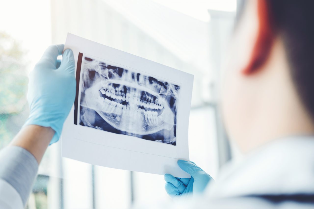
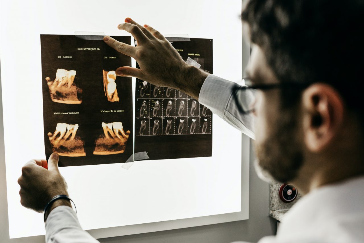
Cone Beam Computed Tomography, literally CBCT, is an abbreviation for cone beam computed tomography, which is a full description of how the structure works. The dental CT scanner produces cone-shaped images as it travels 360 degrees around the patient’s head. The conical shape of the field means that fewer photos are used to scan larger areas without overlapping. The X-ray exposure is therefore orders of magnitude lower.
The resulting images are then stitched together by computer software and finally, with each parameter collected, transformed into a 3-dimensional model. This even reveals hidden problems that we had not anticipated. So the only positive difference with the panoramic X-ray is not just the extremely low radiation exposure. Because digital images reproduce everything to an accuracy of tenths of a millimetre, with no distortion, projection or warping.
The scan itself takes a maximum of one minute and the patient can be comfortable during the process. This is a huge advantage, especially for those who may suffer from claustrophobia: you don’t have to get into a cramped tube. Of course, this is a more comfortable solution for everyone.
To sum up, modern dental CT has brought a new colour to the world of dental treatments, which gives us a complete picture of our dentition and the underlying conditions, excluding errors and revealing everything.
Dental CT imaging is mainly used for implant placement and bone grafting. The more information a surgeon has about the anatomy of a patient’s mouth before placing a dental implant, the better the result. From the image, the surgeon can accurately determine the depth, width, density and condition of the bone, as well as the exact location of the patient’s nerve canals and sinuses. This significantly reduces the patient’s risk and increases the success of the implant.
In the case of dental surgery, there is no need to worry about receiving dangerous levels of radiation, even if, for example, an X-ray has to be redone or several smaller images of separate teeth or implants have to be taken. It should also be noted that, thanks to the rapid advances in technology, X-ray and CT equipment is using lower doses of radiation every year, eliminating unnecessary exposure but still producing sharp, high-quality images.
These imaging techniques are carried out with caution and should only be performed by staff who are qualified and it is in the patient’s interest to receive the desired result after the procedure, in the safest possible conditions, with maximum preparation by the doctors.
